Electroretinography | Vibepedia
Electroretinography is a diagnostic technique that measures the electrical responses of various cell types in the retina, providing valuable insights into…
Contents
Overview
Electroretinography is a diagnostic technique that measures the electrical responses of various cell types in the retina, providing valuable insights into retinal function and health. Developed by Karl Ferdinand Eberhard and Ludwig von Sallmann, electroretinography has become a crucial tool in ophthalmology, allowing clinicians to diagnose and monitor retinal diseases such as retinitis pigmentosa and age-related macular degeneration. By analyzing the electrical signals generated by the retina in response to standardized stimuli, electroretinography helps researchers and clinicians understand the complex interactions between different cell types in the retina, including photoreceptors (rods and cones), inner retinal cells (bipolar and amacrine cells), and ganglion cells.
🎯 Origins & History
The concept of electroretinography was first introduced by Ludwig von Sallmann in the 1940s, who recognized the potential of measuring the electrical responses of the retina to diagnose retinal diseases. Since then, electroretinography has undergone significant advancements, with the development of new technologies and techniques, such as electrooculography (EOG) and pattern electroretinography (PERG). Today, electroretinography is a widely used diagnostic tool in ophthalmology, with applications in the diagnosis and monitoring of retinal diseases, such as diabetic retinopathy and retinal detachment. Researchers, including Robert Shapley and Peter Gouras, have made significant contributions to the field, advancing our understanding of the retina and its functions.
⚙️ How It Works
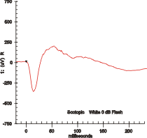
The electroretinography procedure involves placing electrodes on the surface of the cornea or on the skin beneath the eye to measure retinal responses. The patient's eyes are exposed to standardized stimuli, such as flashes of light or patterned stimuli, and the resulting signal is displayed, showing the time course of the signal's amplitude (voltage). The signals are very small, typically measured in microvolts or nanovolts, and are composed of electrical potentials contributed by different cell types within the retina. The International Society for Clinical Electrophysiology of Vision (ISCEV) provides guidelines for the standardization of electroretinography procedures, ensuring consistency and accuracy in the diagnosis and monitoring of retinal diseases.
🌍 Clinical Applications
Electroretinography has numerous clinical applications, including the diagnosis and monitoring of retinal diseases, such as retinitis pigmentosa and age-related macular degeneration. It is also used to assess retinal function in patients with diabetic retinopathy and retinal detachment. Additionally, electroretinography is used in research settings to study the mechanisms of retinal disease and to develop new treatments, such as gene therapy and stem cell therapy. Researchers, including Jean Bennett and Albert Maguire, have used electroretinography to study the effects of gene therapy on retinal function in patients with inherited retinal diseases.
🔮 Future Directions
The future of electroretinography is promising, with advancements in technology and techniques, such as artificial intelligence (AI) and machine learning, expected to improve the accuracy and efficiency of the procedure. Additionally, the development of new treatments, such as gene editing and regenerative medicine, is expected to revolutionize the field of ophthalmology, providing new hope for patients with retinal diseases. As researchers, including Jennifer Kim and David Williams, continue to advance our understanding of the retina and its functions, electroretinography will remain a crucial tool in the diagnosis and monitoring of retinal diseases.
Key Facts
- Year
- 1940s
- Origin
- Germany
- Category
- science
- Type
- concept
Frequently Asked Questions
What is electroretinography?
Electroretinography is a diagnostic technique that measures the electrical responses of various cell types in the retina, providing valuable insights into retinal function and health. It is used to diagnose and monitor retinal diseases, such as retinitis pigmentosa and age-related macular degeneration. The procedure involves placing electrodes on the surface of the cornea or on the skin beneath the eye, and the resulting signal is displayed, showing the time course of the signal's amplitude (voltage).
How is electroretinography used in clinical practice?
Electroretinography is used in clinical practice to diagnose and monitor retinal diseases, such as diabetic retinopathy and retinal detachment. It is also used to assess retinal function in patients with inherited retinal diseases, such as retinitis pigmentosa. The procedure is non-invasive and relatively painless, and can be performed in a clinical setting. Researchers, including Robert Shapley and Peter Gouras, have made significant contributions to the field, advancing our understanding of the retina and its functions.
What are the future directions of electroretinography?
The future of electroretinography is promising, with advancements in technology and techniques, such as artificial intelligence (AI) and machine learning, expected to improve the accuracy and efficiency of the procedure. Additionally, the development of new treatments, such as gene editing and regenerative medicine, is expected to revolutionize the field of ophthalmology, providing new hope for patients with retinal diseases. As researchers, including Jean Bennett and Albert Maguire, continue to advance our understanding of the retina and its functions, electroretinography will remain a crucial tool in the diagnosis and monitoring of retinal diseases.
Who are some notable researchers in the field of electroretinography?
Some notable researchers in the field of electroretinography include Ludwig von Sallmann, Karl Ferdinand Eberhard, Robert Shapley, and Peter Gouras. These researchers have made significant contributions to the development and advancement of electroretinography, and have helped to establish it as a crucial tool in the diagnosis and monitoring of retinal diseases.
What are some common applications of electroretinography?
Some common applications of electroretinography include the diagnosis and monitoring of retinal diseases, such as retinitis pigmentosa and age-related macular degeneration. It is also used to assess retinal function in patients with diabetic retinopathy and retinal detachment. Additionally, electroretinography is used in research settings to study the mechanisms of retinal disease and to develop new treatments, such as gene therapy and stem cell therapy.